Esquizencefalia: Diagnóstico prenatal por medio de ultrasonografía y
Drs. Diego Masoli I(1), Fernando Urzúa V(1), Juan Guillermo Rodríguez A(4), Osvaldo Koller C(2), Mauro Parra C(3), Hernán Muñoz S(3), Oscar Pizarro R(4). 1. Becados de Obstetricia y Ginecología, Campus Oriente, Facultad de Medicina, Universidad de Chile. 2. Instituto de Neurocirugía e Investigaciones Cererales Dr. Alfonso Asenjo. 3. Unidad de Medicina Fetal, Hospital Clínico Universidad de Chile. 4. Centro de Referencia Perinatal Oriente (CERPO), Servicios de Obstetricia y Ginecología y Neonatología, Hospital Santiago Oriente Dr. Luis Tisné Brousse. Abstract Schizencephaly is a rare congenital cerebral malformation; a disorder of neuronal migration characterized by cerebrospinal fluid-filled clefts that extend across the entire cerebral hemisphere, communicating the pial surface with the ventricular surface and lined with gray matter. The clefts may be unilateral or bilateral and may be closed (fused lips), as in schizencephaly type I, or separated (open lips), as in type II. Presentation and outcome are variable, but patients typically present with seizures, hemiparesis and developmental deficits during childhood. There are few reports about the diagnosis of this entity during prenatal life in scientific literature, but they have been increasing due to the development of high resolution ultrasound (US), CAT scan and MRI. We present two cases in which prenatal diagnosis was made, one at 28 and the other at 35 gestational weeks. The diagnosis was made by fetal US and MRI in healthy patients referred to CERPO after it was suspected a cerebral malformation in routine US. We show the US and MRI images of each fetus and a picture of one of the new born. The usefullness of CAT scan and MRI in the diagnosis of this pathology, after the suspicion in the US, must be emphasized. Nonetheless, MRI is the diagnostic method of choice because of its superior differentiation of gray and white matter and its ability to image in more than one plane the fetal anatomy. Key Words: Schizencephaly, Prenatal diagnosis. Resumen La esquizencefalia en una malformación cerebral congénita, de baja incidencia, que se caracteriza por hendiduras en la corteza cerebral comunicando el espacio subaracnoídeo con el sistema ventricular. Se manifiesta en la infancia con alteraciones motoras y convulsiones y en este período se realiza la mayoría de los diagnósticos. Los reportes de diagnóstico en la vida fetal son escasos, pero han aumentado desde el advenimiento de la ultrasonografía de alta resolución (US), la tomografía computada (TC) y la resonancia magnética (RM). Presentamos dos casos en los que se realizó diagnóstico prenatal; a las 28 y 35 semanas de gestación. El diagnóstico se realizó mediante US y RM fetal, en mujeres embarazadas sanas referidas al CERPO por malformación cerebral en US de rutina. Se muestran imágenes de US y RM de ambos fetos y de uno de los recién nacidos. Destacamos la utilidad de la TC o RM para efectuar el diagnóstico de certeza cuando se ha sospechado esta malformación a la US asimismo para efectuar el diagnóstico diferencial con otras entidades como la holoprosencefalia, hidrocefalia y porencefalia, poniendo énfasis en la RM por su mejor resolución y su despliegue multiplanar de la anatomía fetal. Palabras Claves: Esquizencefalia, Diagnóstico prenatal. Resonancia Magnética prenatal demuestra imagen de malformación cerebral. Resonancia magnética prenatal demuestra imagen de defecto intracerebral. Masoli D. y cols. Esquizencefalia: Diagnóstico prenatal por medio de ultrasonografía... Rev Chil Ultrasonog 2004; 7:40-44. Correspondencia: Dr. Dego Masoli diegomasoli@yahoo.es Polipectomía endometrial simplificada sin histeroscopía quirúrgica Volver al sumario volumen 7, número 2
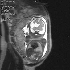
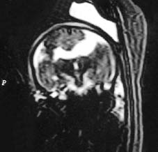
Resonancia Magnética prenatal demuestra imagen de malformación cerebral. Resonancia magnética prenatal demuestra imagen de defecto intracerebral.
Polipectomía endometrial simplificada sin histeroscopía quirúrgica Volver al sumario volumen 7, número 2
Drs. Diego Masoli I(1), Fernando Urzúa V(1), Juan Guillermo Rodríguez A(4), Osvaldo Koller C(2), Mauro Parra C(3), Hernán Muñoz S(3), Oscar Pizarro R(4).
1. Becados de Obstetricia y Ginecología, Campus Oriente, Facultad de Medicina, Universidad de Chile.
2. Instituto de Neurocirugía e Investigaciones Cererales Dr. Alfonso Asenjo.
3. Unidad de Medicina Fetal, Hospital Clínico Universidad de Chile.
4. Centro de Referencia Perinatal Oriente (CERPO), Servicios de Obstetricia y Ginecología y Neonatología, Hospital Santiago Oriente Dr. Luis Tisné Brousse.
Abstract
Schizencephaly is a rare congenital cerebral malformation; a disorder of neuronal migration characterized by cerebrospinal fluid-filled clefts that extend across the entire cerebral hemisphere, communicating the pial surface with the ventricular surface and lined with gray matter. The clefts may be unilateral or bilateral and may be closed (fused lips), as in schizencephaly type I, or separated (open lips), as in type II. Presentation and outcome are variable, but patients typically present with seizures, hemiparesis and developmental deficits during childhood. There are few reports about the diagnosis of this entity during prenatal life in scientific literature, but they have been increasing due to the development of high resolution ultrasound (US), CAT scan and MRI. We present two cases in which prenatal diagnosis was made, one at 28 and the other at 35 gestational weeks. The diagnosis was made by fetal US and MRI in healthy patients referred to CERPO after it was suspected a cerebral malformation in routine US. We show the US and MRI images of each fetus and a picture of one of the new born. The usefullness of CAT scan and MRI in the diagnosis of this pathology, after the suspicion in the US, must be emphasized. Nonetheless, MRI is the diagnostic method of choice because of its superior differentiation of gray and white matter and its ability to image in more than one plane the fetal anatomy.
Key Words: Schizencephaly, Prenatal diagnosis.
Resumen
La esquizencefalia en una malformación cerebral congénita, de baja incidencia, que se caracteriza por hendiduras en la corteza cerebral comunicando el espacio subaracnoídeo con el sistema ventricular. Se manifiesta en la infancia con alteraciones motoras y convulsiones y en este período se realiza la mayoría de los diagnósticos. Los reportes de diagnóstico en la vida fetal son escasos, pero han aumentado desde el advenimiento de la ultrasonografía de alta resolución (US), la tomografía computada (TC) y la resonancia magnética (RM). Presentamos dos casos en los que se realizó diagnóstico prenatal; a las 28 y 35 semanas de gestación. El diagnóstico se realizó mediante US y RM fetal, en mujeres embarazadas sanas referidas al CERPO por malformación cerebral en US de rutina. Se muestran imágenes de US y RM de ambos fetos y de uno de los recién nacidos. Destacamos la utilidad de la TC o RM para efectuar el diagnóstico de certeza cuando se ha sospechado esta malformación a la US asimismo para efectuar el diagnóstico diferencial con otras entidades como la holoprosencefalia, hidrocefalia y porencefalia, poniendo énfasis en la RM por su mejor resolución y su despliegue multiplanar de la anatomía fetal.
Palabras Claves: Esquizencefalia, Diagnóstico prenatal.
Resonancia Magnética prenatal demuestra imagen de malformación cerebral. Resonancia magnética prenatal demuestra imagen de defecto intracerebral.
Masoli D. y cols. Esquizencefalia: Diagnóstico prenatal por medio de ultrasonografía... Rev Chil Ultrasonog 2004; 7:40-44.
Correspondencia: Dr. Dego Masoli
diegomasoli@yahoo.es
Polipectomía endometrial simplificada sin histeroscopía quirúrgica Volver al sumario volumen 7, número 2